제주 해안주변해역 표층퇴적물 중 와편모조류 시스트 군집의 분포특성
초록
제주 해안선 주변 항만 표층퇴적물의 와편모조류 시스트 군집구조를 파악하기 위하여, 제주 해안을 따라 위치하는 22곳의 항만을 대상으로 2012년에서 2016년까지 격년단위로 3회 조사하였다. 조사는 소형 에크만 채니기를 이용하여 0~2 cm 두께의 표층퇴적물 표본을 채집하였다. 분석결과 출현이 확인된 와편모조류 시스트는 6그룹 9속 29종으로 Protoperidinioid Group이 13종으로 44.8%의 점유율을, Gonyaulacoid Group이 9종으로 31.0%, Calciodineloid Group이 3종으로 10.3%, Gymnodinioid Group이 2종으로 6.9%, Tuberculodinoid Group과 Diplosalid Group이 각각 1종으로 3.5%의 점유율을 보였다. 또한 정점에 따른 변화는 1~6 종으로 매우 낮았다. 시스트의 세포밀도는 13~220 cysts/g dry의 범위로 낮지만, 퇴적물의 함수율과 유의적인 회귀식을 나타내었다. 또한 일부 세립퇴적물을 나타내는 항만 및 개발에 의해 인위적으로 형성된 조수웅덩이에서는 종속영양종 출현비가 높아, 오랜 시간 유기물이 퇴적되고 있음을 시사하였다. 우점종은 cyst of Gymnodinium sp 및 Protoperidinium 속 시스트가 최우점하였고, 이외 cyst of Pyrodinium bahamense, cyst of Scrippsiella trochoidea 등이 우점 출현하였다. 특히 열대 및 아열대성 유독와편모조인 P. bahamense 시스트는 제주연안에서 처음 기록되는 종으로 제주 해안주변해역의 인위적 조수웅덩이 등의 해역 수질환경 및 열대나 아열대에서 유입되는 새로운 유독와편모조류에 대한 모니터링 및 관리방안 수립이 시급한 것으로 판단되었다.
Abstract
This study describes the spatial distribution of dinoflagellate cyst assemblages from the fishing ports along Jeju Island. Surface sediment samples from 22 stations revealed the occurrence of 29 species involving the Groups Protoperidinioid (44.8%), Gonyaulacoid (31.0%), Calciodineloid (10.3%), Gymnodinioid (6.9%), Diplosalid (3.5%) and Tuberculodinioid (3.5%). The cyst abundance recorded here is very low (13~220 cysts g-dry-1) as compared to Korean coastal regions. The abundance of heterothophic cysts increased in several fishing pots with fine sediments and anthropogenic tidal pools. And cyst abundance was well correlated with the grain-size composition of surface sediments. The dinoflagellate cyst assemblages in Jeju fishing ports were characterized by the dominant species, cyst of Gymnodinium sp., cyst of Pyrodinium bahamense and cyst of Scrippsiella trochoidea in 2012, Protoperidinium sp. (Brigantedinium sp.), cyst of Scrippsiella sp./trochoidea and cyst of Gymnodinium sp. in 2014, and Protoperidinium sp. (Echinidinium sp. and Brigantedinium sp.) in 2016. The advent of the toxic dinoflagellate, Pyrodinium bahamense were recorded for the first time in Jeju coastal waters. As a results, we are determined should be to monitoring and management measures for new toxic dinoflegallates from tropical or subtropical reigions and anthropogenic tidal pools by industrial activities.
Keywords:
Dinoflagellate cysts, cell density, autotrophic and heterotrophic dinoflagellates, dominant species, Jeju fishing ports, Pyrodinium bahamense, anthropogenic tidal pool키워드:
와편모조류 시스트, 세포밀도, 독립 및 종속와편모조류, 우점종, 제주 항만, 피로디니움 바하멘스, 인위적 조수웅덩이1. 서 론
와편모조류는 해양생태계에서 기초생산을 담당하는 식물플랑크톤 군집의 중요한 구성원으로 규조류 다음의 출현종과 현존량을 나타낸다. 현재 약 2,500종의 와편모조류가 보고되고 있으며(Taylor[1987]; Anderson et al.[1998]; Goméz[2012]), 이들 분류군은 단세포로서 일반적인 식물플랑크톤과 같이 표영환경에서 부유생활을 할 때에는 무성생식을 한다. 그러나 서식환경이 악화되면 배우자와 접합하여 내구성 휴면접합자인 휴면포자, 즉 시스트를 만들어 해저에 침강하여 서식에 부적합한 환경을 지내고, 재차 서식환경이 좋아지면 발아하여 부유생활을 하는 생활사를 가진다(Anderson et al. [1983]; Walk[1984]). 현재 이러한 생활사에서 휴면포자인 시스트를 만드는 와편모조류는 전체 보고되는 종의 약 10% 정도 알려진다(Mastuoka and Fukuyo[ 2000]; Bravo and Figueroa[2014]). 이러한 와편모조류 시스트의 생활사적 특성은 표영환경을 나타내는 누적지표로서 연안해역의 생물해양학적 환경특성 파악에 중요한 지표로 활용된다(Yoon and Shin[2014]). 또한 와편모조류는 광합성을 하는 기초생산자이나, 실제로 완전한 독립영양종은 몇 종에 지나지 않고, 전체에서 약 절반은 혼합영양, 그리고 나머지 절반은 종속영양을 하는 등 다양한 영양방식을 구사한다(Gains and Elbrächter[1987]. 때문에 이들 종속영양종과 독립영양종 시스트의 출현비를 이용하여 연안해역의 부영양화과정 등 해양환경의 변화과정을 추적하는 유효한 방법으로 사용되기도 한다(Dale et al.[1999]; Matsuoka[1999]; Dale[2001], [2009]; Ismael et al.[2014]).
한국 연안해역에서 시스트 연구는 1990년 이후의 일(Kim et al.[1990])로서 2000년 이후 본격적으로 수행하기 시작하였다(Yoon and Shin[2013]. 그러나 연구 대부분은 남해의 내만 및 연안해역을 대상으로 한 것(Lee and Yoo[1991]; Lee and Matsuoka[1996]; Lee et al.[1998]; Cho et al.[2003]; Park and Yoon[2003]; Park et al.[2005]; Shin et al.[2007]; Kim et al.[2009]; Pospelova and Kim[2010]; Shin et al.[2011])이며, 서해 및 황해(Cho and Matsuoka[2001]; Park et al.[2004]; Hwang et al.[2009], [2011]), 그리고 제주도를 포함한 동중국해(Cho and Matsuoka[2001]; Cho et al.[2004])를 대상으로 한 일부 결과가 있다. 특히 이 논문에서 연구대상이 되는 제주도는 화산도로서 해안선 대부분이 암반 또는 모래해안으로 구성되고 있어, 시스트 연구를 위한 해저퇴적물 채집이 매우 어렵거나 거의 불가하다. 단지 해안선의 자연부락에 어선의 정박이나 피항에 대비한 소형지방항만과 국가항만에서 세립한 퇴적물이 존재하고 있을 뿐이다. 때문에 제주연안에서는 아직까지 인간생활의 영향을 받는 천해역에서 수행된 와편모조류 시스트 연구는 찾을 수 없다.
따라서 이 연구는 제주도 해안선의 항만을 대상으로 표층퇴적물 중의 와편모조류 시스트의 분포특성을 이용하여, 제주 주변 천해역의 해양환경실테 파악은 물론 생물해양학적 특성을 해석하여, 앞으로 제주 연안천해역의 이용과 관리, 그리고 환경보전 대책 수립에 필요한 자료를 제공하고자 한다.
2. 재료 및 방법
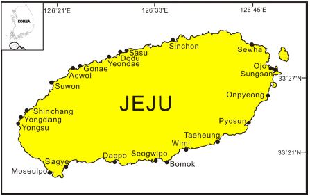
제주도 해안선을 따라 표층퇴적물 채집이 가능했던 항만을 대상으로 현장조사를 실시하였다. 즉 조사는 제주 해안선 주변 대부분 항만을 대상으로 하였지만, 해저저질 구성이 암반, 자갈 및 조립한 사질인 곳을 제외하고, 2012년 9월 18곳, 2014년 11월 9곳 그리고 2016년 6월 16곳의 항만에서 표층퇴적물 채집이 가능하였다(Fig. 1). 채집은 주로 와편모조류 출현이 많은 여름과 가을을 대상으로 실시하였다. 표층퇴적물은 항만의 시설물 또는 어선을 계류할 수 있게 조성된 제방에서 표준 에크만 채니기(Ekman-Birge, Hydro-Bios No.437-200)를 이용하여 채집하였다. 채집된 표본은 현장에서 디지털 온도계(Electronic Temperature Instruments Ltd (ETI), 231-257)를 이용하여 표층퇴적물 온도 측정과 함께 플라스틱 스푼으로 0~2 cm의 표층퇴적물을 밀폐할 수 있는 소형 플라스트 용기에 채집하였다. 채집된 표본은 아이스박스를 이용하여 실험실로 운반한 다음 냉장 보관하여 검경 표본을 준비하였다. 시스트 채집과 함께 잠수형형광광도계(JFE Advantech Co., Ltd, ASTD 102)를 이용하여 현장에서 저층 해수의 수온과 Chlorophyll a (Chl-a) 농도를 측정하였다.
시스트 동정 및 계수를 위한 검경시료는 퇴적물 1 g를 원심관에 채취하여 증류수 10 mL를 첨가하여 원심분리기를 이용 염분을 제거하였다. 퇴적물의 염분을 완전히 제거하기 위해 이 과정을 2~3회 반복한다. 탈염된 표본에 10% HCl 5 mL 첨가하여 후드에서 약 24시간 방치하여 미세플랑크톤이나 방상충 등을 제거하였다. 제거된 표본은 증류수를 이용하여 2~3회 세척을 한다. 세척된 표본은 5% Ca(OH)2 용액 5 ml를 첨가하여 약 70 oC에서 3분간 중탕하여 유기물질을 제거하였다. 동일한 세척과정으로 Ca(OH)2 용액을 제거한 다음 25~30%의 hydrofluoric acid 5~10 mL를 첨가하여 약 70 oC에서 2~3시간 중탕하여 규산질 입자를 제거한 다음 세척하였다. 이렇게 준비된 표본을 초음파분쇄기(Bandelin Electronic, UW2070)로 분쇄한 다음 망목이 125 μm와 20 μm 체를 중첩하여 시스트를 세척하면서 농축하여, 20 μm 체에 모여진 물질을 샬렛에 옮겨 미세사질과 시스트의 비중 차를 이용하여 미세사질을 제거하였다(Matsuoka and Fukuyo[2000]; Yoon and Shin[2014]). 이렇게 준비된 표본은 10 mL로 정용하여 검경표본으로 하였다. 종의 동정과 계수는 검경표본 1 mL를 정확하게 Sedgwick-Rafter Chamber에 취하여 광학현미경(Olymps BX50, Nikon Eclipse 80i)을 이용하여 검경을 실시하였고, 시스트의 현존량은 단위 건중량당 세포수(cysts/g dry)로 나타내었다((Matsuoka and Fukuyo[2000]; Yoon and Shin[2013]).
그리고 현존량 및 유기물 및 시스트의 집적 정도를 간접적으로 파악하기 위한 함수율은 채집된 퇴적물 약 5 g를 도가니에 넣고 퇴적물 취해 건조기를 이용 110 oC에서 24시간 건조하여 상온으로 내린 다음 건조 전, 후의 무게를 이용한 식, 즉 Water content (%) ={(wet weight - dry weight) / wet weight} × 100(%)으로 계산하였다(OSC[1986]). 또한 얻어진 2016년의 환경자료 및 우점종 자료는 SPSS 프로그램을 이용한 주성분분석(PCA)으로 와편모시스트 출현 특성을 파악하였다.
3. 결 과
3.1 와편모조류 시스트 군집
제주도 해안선 주변 항만 표층퇴적물에서 확인된 와편모조류 시스트는 시스트명을 기준으로 6그룹 9속 29종이였고, 플랑크톤 종 기준으로는 9속 27종이 출현하였다(Plate 1 and Plate 2). 그룹별로는 Protoperidinoid group이 13종으로 44.8%의 점유율을, Gonyaulacoid group이 9종으로 31.0%, Calciodineloid group이 3종으로 10.3%, Gymnodinioid group이 2종으로 6.9%, 그리고 Tuberculodinoid group과 Diplosalid group이 각각 1종으로 각 3.5%의 점유율을 나타내었다. 년도별로는 2012년에 5그룹 17종으로 Protoperidinioid group이 7종에 41.2%, Gonyaulacoid group이 4종에 23.5%, Calciodineloid group이 3종에 17.6%, Gymnodinioid group이 2종에 11.8% 그리고 Tuberculodinoid group이 1종으로 5.9%의 점유율을 나타내었다. 2014년은 5그룹 13종으로 Protoperidinioid group과 Gonyaulacoid group이 각 4종으로 각 30.8%, Gymnodinioid group이 2종으로 15.4%, Calciodineloid goup이 2종으로 15.4%, 그리고 Diplosalid group이 1종으로 7.6%의 점유률을 나타내었다. 2016년은 5그룹 17종으로 Protoperidinioid group이 9종에 52.9%, Gonyaulacoid group이 4종에 23.5%, Gymnodinioid group이 2종에 11.8%, 그리고 Calciodineloid group과 Tuberculodinoid group이 각 1종으로 5.9%의 점유율을 나타내었다. 그리고 영양방법에 따른 와편모조류 시스트의 출현비는 독립영양종이 15종으로 51.7%, 종속영양종이 14종으로 48.3%를 차지하였다(Table 1).
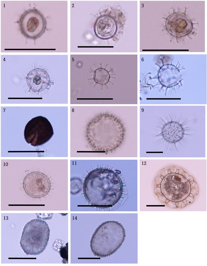
Photomicrographs of autotrophic dinoflagellate cysts recorded from Jeju Island. 1: Gonyaulax digitalis (Spiniferites bentorii), 2: Gonyaulax sp.1 (Spiniferites hypercanthus), 3: Gonyaulax sp2. (Spiniferites delicatus), 4: Gonyaulax membranaceus (Spiniferites membranaceus), 5: Gonyaulax scrippsae (Spiniferites bulloideus), 6: Gonyaulax verior, 7: Gymnodinium catenatum, 8: Lingulodinium polyedrum (Lingulodinium machaerophorum), 9: Lingulodinium machaerophorum (Lingulodinium machaerophorum) 10: Protoceratium reticulatum (Operculodinium centrocarpum) 11: Pyrodinium bahamnese (Polysphaeridinium zoharyi), 12: Pyrophacus steinii (Tuberculodinium vancampoae), 13: Scrippsiella sp., 14. Scrippsiella sp. cf. trochoidea. The name in ( ) showing palaeontological name. (scale bar : 50 μm).
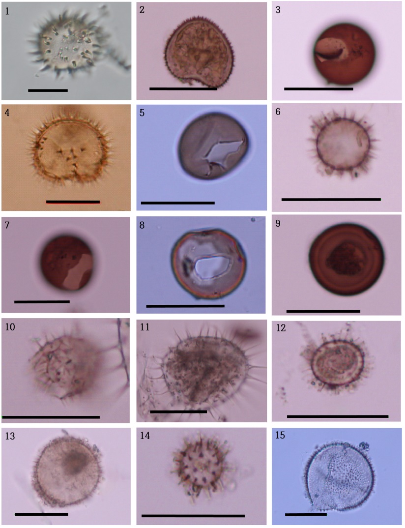
Photomicrographs of heterotrophic dinoflagellate cysts recorfed from Jeju Island. 1: Oblea acanthocysta (Oblea acanthocysta), 2: Protoperidinium claudicans (Votadinium spinosum), 3: Protoperidinium conicoides (Brigantedinium simplex), 4: Protoperidinium conicum. (Selenopemphix quanta), 5: Protoperidinium denticulatum (Brigantedinium irregulare), 6: Protoperidnium minutum, 7: Protoperidinium sp.1 (Brigantedinium asymmetricum), 8: Protopeidinium sp. 2 (Brigantedinium sp. 1), 9: Protoperidinium sp. 2. (Brigantedinium sp. 2), 10: Protoperidinium sp. 3 (Echinidinium delicatum), 11: Protoperidinium sp. 4 (Echinidinium granulatum), 12: Protoperidinium sp. 5 (Echinidinium sp.), 13: Protoperidinium sp. 6 (Islandinium brevispinosum), 14: Protoperidinium sp. 7 (Islandinium minutum), 15: Protopeidinium sp. 8 (Islandinium sp.). The name in ( ) showing palaeontological name. (scale bar : 50 μm).
와편모조류 시스트 출현 종의 시·공간분포는 2014년 13종, 2012년과 2016년이 각 17종이었지만, 정점별로는 1~6 종으로 매우 낮았다. 3회 조사에서 제주 북쪽 바다를 면하는 도두항과 세화항에서 출현종수가 많았고, 남쪽 바다를 면하는 사계항과 위미항에서 낮았다. 2012년에는 1~5 종의 출현종이 나타났다. 그중 서귀포항에서 5종으로 가장 많은 출현 종수를 나타냈으며, 위미항과 고내항에서는 1종으로 가장 낮은 출현 종수를 나타내었다. 2014년에는 1~6 종의 출현종이 나타났다. 그중 세화항에서 가장 많은 출현종수를 보였으며, 용수항에서 가장 낮은 출현 종수를 나타내었다. 2016년에는 1~4 종의 출현종이 나타났다. 그중 도두항과 세화항 모슬포항에서 4종으로 높은 출현 종수를 나타내었으며 사수항과 오조항, 위미항에서 1종으로 낮은 출현 종수를 나타내었다(Fig. 2).
제주주변 항만 표층퇴적물에서 관찰된 와편모조류 시스트 현존량은 전체 13~220 cysts/g dry의 범위로 매우 낮은 값을 보였다. 조사시점에 따라서도 2012년 23~198 cysts/g dry, 2014년은 16~165 cysts/g dry, 그리고 2016년은 13~220 cysts/g dry의 출현 범위로 조사시점에 따른 변동은 크지 않았다(Fig. 3). 그리고 출현한 와편모조류를 영양방법에 의해 독립영양종과 종속영양종으로 구분하여 계산하면, 2012년은 독립영양종보다 종속영양종이 각 78.4% 및 21.6%로 독립영양종이 출현 점유률이 높았다. 같은 방법으로 2014년은 63.3% vs 36.7%로 2012년과 유사하였지만, 2016년은 29.8% vs 70.2%로 종속영양종이 더 높은 비율로 출현하여 2012년 및 2014년과는 상반되는 경향을 나타내었다(Fig. 3).
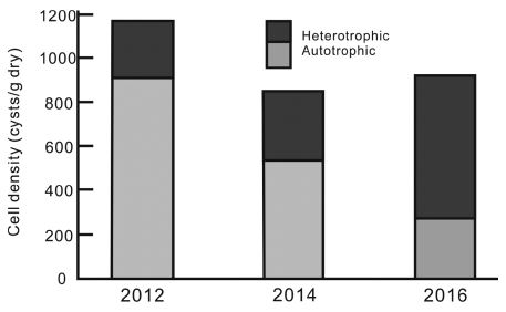
Bi-yearly change of dinoflagellate cyst density (invoving densities of autotrophic and heterotrophic dinoflagellte cysts).
시스트 현존량의 공간분포에서 2012년은 항만 위치에 대한 규칙성은 관찰되지 않았지만, 남쪽 위미항에서 낮았고, 북동쪽 신촌항에서 높았다. 2014년은 2012년과는 달리 서쪽의 용수항에서 낮았고, 북쪽 제주공항에 인접한 도두항에서 높았다. 그리고 2016년은 용두암 인근에 위치한 사수항에서 낮은 반면, 북동해역에 위치하는 세화항에서 높은 현존량을 보였다(Fig. 4). 영양방법에 따른 출현양상은 2012년의 경우, 제주 동부의 세화항, 상산항 및 남부의 보목항과 태흥한, 그리고 서부의 신창항을 제외하면 모두 독립영양종에 의해 출현이 점유되었다. 동쪽과 남부 일부 지역에서는 종속영양종의 비율이 독립영양종의 비율 보다 높게 나타났다. 2014년은 서부의 용수항에서 낮았고, 제주공항 인근인 도두항에서 높았다. 영양방법에 따라서는 용수항, 도두항, 사계항, 그리고 대포항 등 서쪽해역에서 종속영양종 출현이 높았다. 2016년은 독립영양종이 사수항에서 낮고, 세화항에서 높은 세포밀도를 나타내었다. 그리고 신촌항 및 오조항을 제외한 나머지 정점에서 종속영양종이 높은 비율로 출현하였다(Fig. 4).
제주 해안의 표층퇴적물에 출현한 와편모조류 시스트 중 전체 정점 평균으로 10% 이상 우점율을 나타내는 종은, 2012년은 cyst of Gymnodinium sp, cyst of Pyrodinium bahamannese 및 cyst of Scrippsiella trochoidea로서 모두 독립영양종에 의해 우점되었다. 2014년 역시 cyst of Protoperidinium sp. (Brigantedinium sp.), cyst of Scrippsiella sp., cyst of Gymnodinium sp. 그리고 cyst of Scrippsiella trochoidea가 10% 이상 우점율을 보여 cyst of Protoperidinium sp.의 22.5%의 우점율을 제외하면 31.9%가 독립영양종에 의해 우점되었다. 그러나 2016년은 Protoperidinium sp. 5 (Echinidinium sp.) 및 Protoperidinium sp. 2 (Brigantedinium sp.)등 Protoperidinium 속의 우점율이 46.2%로 2012, 2014년과는 달리 종속영양성 와편모조류 시스트가 높게 나타나는 특징을 보였다(Table 2).
제주도는 화산도로서 대부분 해안선은 암반 및 일부 모래해안으로 구성되어 있으나, 소형 어선의 계류 및 피항 시설이 되어 있는 소형항만은 외부 파랑의 유입정도에 따라 일부 해역에 뻘이 퇴적되어 와편모조류 시스트 등 표영환경에서 침강된 생물기원물질이 퇴적될 수 있는 기반이 조성되어 있다. 이러한 해역에서 유기물 등 표영환경의 다양한 물질의 퇴적가능 정도를 간접적으로 파악할 수 있는 지표가 함수율이다. 제주주변의 항만에서 함수율은 14.2~75.8%로 큰 변동 폭을 나타내었다. 시·공간적으로 다소 차이는 있지만 남부의 보목항 및 사계항 이나 북부의 도두항, 신총항 및 세화항 등 2중 방파제에 의해 해수교환이 원활하지 않은 항내의 안쪽 정점에서 높은 값을 보였고, 항외 해수가 쉽게 유입될 수 있는 남부의 위미항 및 북부의 사수항 등에서 낮은 함수율을 나타내었다(Fig. 5). 동일 항만에서 년도에 따른 차이점은 표층퇴적물 채집위치가 다소 다르기 때문에 나타나는 현상이다.
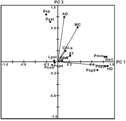
2016년 각 항만 평균 3% 이상의 우점율을 가지는 와편모조류 시스트 및 퇴적물표층의 온도 및 함수율, 그리고 저층해수의 염분 및 Chl-a 농도 등의 환경인자를 이용한 주성분분석을 누적기여율 70%로 계산하면 Z = 4.510Z1 + 2.728Z2 + 2.312Z3 + 1.960Z4가 되어 제4주성분까지 도출되지만, 제3주성분까지 누적기여율이 63.7%를 나타내고 있어, 제3주성분까지 결과를 Table 3에 정리하였다. 그러나 출현종에 의한 주성분분석에서 각 종에 대한 특성 파악이 어렵기에 편의상 여기서는 제2주성분, 즉 누적기여율 48.3%까지를 대상으로 검토하였다.

Eigen value, proportion, eigen vector and loading factor by PCA on the dinoflagellate cysts around Jeju Island in 2016
인자부하량에 의해 와편모조류 시스트의 분포 양상은 제1주성분에는 종속영양종 시스트, 제2주성분에는 독립영양종 시스트가 집중적으로 분포하였다. 인자부하량의 분포에서 독립영양종과 종속영양종 사이의 상관은 낮은 것으로 나타났지만, 종속영양종 전체(HD) 및 우점종은 저층 해수의 염분과 퇴적물온도에 높은 상관성을 가지는 것으로 나타났다. 그러나 독립영양종 중 Gonyaulax scrippsea는 종속영양종의 분포특성과 유사한 출현특성을 나타내고 있었으며, 환경인자 중 함수율과 Chl-a 농도는 독립영양종 전체(AD)와는 강한 상관을 보이지만, 우점종과의 사이에는 종속영양종 유사한 관련성을 나타내었다. 그리고 함수율과 저층 해수의 Chl-a 농도는 거의 일직선으로 분포하고 있어, 두 항목은 매우 밀접한 관련성을 가지는 것으로 나타났다(Fig. 6).
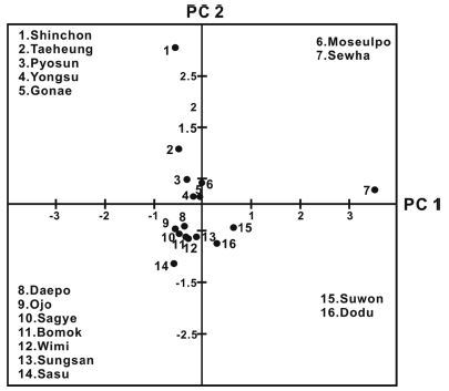
주성분분석에 의한 득점분포에서는 제1주성분에 양의 관계를 보이는 정점으로는 세화, 수원, 도두, 모슬포항이 분포하였고, 제2주성분에는 신촌, 태흥, 표선, 용수 및 고내항이 위치하였다. 기타 대포, 오조, 사계, 보목, 위미, 성산 및 사수항은 제1 및 제2주성분 모두에 음의 관계를 나타내었다(Fig. 7). 제1 및 제2주성분에 모두 음의 관계를 보이는 7개 항만의 특징은 주성분분석에 사용한 우점종 와편모조류가 출현하지 않았거나, 출현 세포밀도가 매우 낮은 정점이었다.
4. 고 찰
한국 연안을 대상으로 연구한 와편모조류 시스트 관련 기존의 46편 문헌에 1회라도 출현하는 것으로 알려진 시스트는 약 90종(Yoon and Shin[2013])이다. 그러나 연구초기인 1990년대 초반에는 10종 내외로 제한적인 종만 동정 되었다(Kim et al.[1990]; Lee and Yoo[1991]. 그리고 현재까지 가장 많은 종은 통영 주변의 양식장을 대상으로 한 연구에서 47종이 보고된 것이다(Pospelova and Kim[2010]). 이러한 결과를 고려하면 제주연안에서 출현한 시스트 30종은 적지 않은 수준이다(Table 4). 그러나 세부적으로 보면 각 정점에 따른 출현종은 1~6종 수준으로 매우 낮았다. 이렇게 정점별 출현종이 낮은 것에 비해 전체 시스트 출현종이 다양한 것은 제주도 주변해역은 계절에 따라 쓰시마난류, 양자강 희석수, 황해저층수괴 등 다양한 수괴의 영향범위가 서로 다르게 나타나는 복잡한 해황특성을 보이기 때문(Lie and Cho[2002])인 것으로 판단할 수 있었다.

The records of dinoflagellate cysts abundance and species number from the Korean coastal waters and Indian ocean
그러나 제주연안의 와편모조류 시스트 현존량은 13~220 cysts/g dry으로 매우 낮다. 고생물학적 연구를 포함하여 지구규모에서 보면 와편모조 시스트의 분포와 현존량은 해역과 정점에 따라 단위 건중량 당 시스트 세포수가 0 ~ 수십만 이상으로(Head[1996]; Zonneveld et al.[2013]), 변동 폭이 큰 특징을 보인다. 국내에서도 동중국해 북부에서 0 cyst/g dry(Cho and Matsuoka[2001])에서 황해 중앙부의 20,828 cyst/g dry(Hwang et al.[2011])까지 큰 변동 폭을 보이나, 대부분 내만에서는 수십에서 수천 cyst/g dry의 범위로 출현하여, 제주 연안의 시스트 현존량은 매우 낮음을 알 수 있다. 그렇지만 최근 인도양의 열대 및 온대해역의 연구결과에서는 수십에서 수백 cyst/g dry의 범위로 제주연안과 큰 차이가 없는 보고도 보인다(Table 4). 해역에 따른 시스트 현존량의 차이는 시스트의 생산과 해수 및 퇴적물 특징에 의해 결정되는 것이 알려지지만(Anderson et al.[1995]; Dale[1983]; Joyce et al.[2005]), 제주연안은 표층퇴적물의 퇴적상과 깊게 관계는 것으로 판단할 수 있었다(Narale et al.[2013]). 제주도는 화산도로서 세립질 퇴적물은 포구 속의 극히 일부해역에서만 채집되고 있을 뿐만 아니라 퇴적상이 차단되어 대부분의 포구의 입구부나 중앙부에는 암반이나 사질퇴적물로 구성되는 특징을 가진다. 그리고 항만 1/2정도에서는 퇴적물 재집이 되지 않았다. 즉 포구가 조성되어 오랜 시간 부유물질이 퇴적되는 극히 일부에서만 시스트 관찰이 가능한 것이 된다. 실제 2012년부터 3회 조사한 퇴적물 함수율과 시스트 현존량 사이에는 강한 양의 상관을 나타내고 있으며, 이 자료에서는 조립한 사질이나 자갈 퇴적상은 포함되지 않았다(Fig. 8).
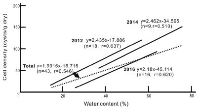
Regression equations between dinoflagellate cyst abundance and water content around Jeju Island in 2012, 2014 and 2016.
최근 와편모조류 시스트의 영양방법을 이용하여 청정해역은 독립 영양종이 우세한 반면, 산업활동이나 저염분에 의한 높은 영양염류 유입으로 규조류에 의한 기초생산이 높아진 부영양화 해역은 종속 영양 와편모조류 출현비가 높다는 내용(Sætre et al.[1997]; Matsuoka[1999], [2001], Pospelova and Kim[2010]; Ismael et al.[2014])을 근거로 와편모조류 시스트를 해역의 부영양화 지표로 활용되고 있다(Dale et al.[1999]; Dale[2001], [2009]). 이러한 내용에 의하면 제주연안은 전체적으로 독립영양종 출현비가 높았던 2012년과 2014년에 비해 2016년은 종속영양종 출현비가 증가하여(Fig. 3), 2016년 부영양화가 급격히 진행된 것이 된다. 그러나 2012년 세화항외 4개항, 2014년 도두항외 2개항, 그리고 2016년 사수항외 10개항에서 독립영양종보다 종속영양종 출현비가 높지만(Fig. 4), 이들을 세부적으로 보면 동일 항만에서도 조사년도에 따라 다른 결과를 나타내고 있으며, 자료로 제시되지는 않았지만, 세화, 태흥, 위미(2016), 보목, 사계, 모슬포(2016), 용수, 신창(2014); 수원(2016), 고내에서 ( )속에 년도 표시가 없는 곳은 3회 모두 세립질퇴적물 채집이 가능한 곳이였다. 그리고 ( )속 연도 표시가 된 곳은 퇴적물 채집이 어려워 해안선에서 다소 격리된 조수웅덩이 표본이었다. 2016년 사수항과 2014년 위미항은 사질퇴적상으로 1종만이 출현하였다. 또한 동일 장소에 대해서도 세립한 퇴적물 채집이 어려워 조사시점마다 채집위치가 다른 것에 기인한다. 즉 이러한 결과로부터 제부 주변해역은 단순한 종속영양 및 독립영양종의 출현비를 이용하여 부영양화 등 환경변화를 추정하는 것은 객관성 확보가 어렵다는 것을 나타낸다. 그러나 제주 해안선 주변 항만은 개방된 해역과는 달리 많은 적조생 물종이 출현 등이 보고되고 있어, 환경문제의 발생지로서 역할을 할 가능성이 지적되고 있다(Yoon et al.[1991]). 이러한 내용과 이번 조사결과를 종합적으로 살펴보면 제주 해안선은 관광수요 증가로 인하여 해안도로 등을 건설하면서 자연해안선 일부가 도로에 의해 격리되어 해수유동이 차단된 인위적 조수웅덩이 등의 해역의 표층퇴적물은 점차 환원상태로 진행되고 있는 것으로 판단되었다. 이와 같은 사실은 추가적인 퇴적물 중의 유기물량이나 황화수소 또는 화학적산소요구량 등 직접적 환경인자 측정에 의한 확인이 필요하지만, 채집된 퇴적물의 검정색으로 변하여. 일부에서 황화물 냄새를 발생하는 것으로도 확인이 가능하였다. 즉, 이는 해안도로나 항만건설로 일부 격리된 크고 작은 조수웅덩이 형태의 해역관리가 시급하게 필요한 것을 시사하는 내용이라 할 수 있을 것이다.
제주 연안의 우점종에서 Gymnodinium sp. 는 진해 및 마산만(Lee et al.[1998]), Protoperidinium group는 가막만(Lee et al.[1999]; Park and Yoon[2003]; Shin et al.[2011], 여자만 및 봇돌바다, 그리고 남해 서부해역(Shin et al.[2007], [2011]), Scrippdiella trochoidea 및 Scrippsiella sp.는 남해의 통영연안, 중앙부 해역, 그리고 서부해역 등 광범위한 해역(Kang et al.[1999]; Park et al.[2005], [2006])에서 우점종으로 출현한 기록이 있는 국내의 보편적 와편모조류 시스트라고 할 수 있다. 그러나 남해안에서 특징적으로 출현하는 Alexandrium속(Shin et al.[2011])이나 Gonyaulax 속(Pospelova and Kim[2010])등은 우점 출현하지 않았다. 그러나 2012년 우점종으로, 2016년에도 출현한 유독플랑크톤, Pyrodinium bahamense는 한국연안에서 남해 통영연안이나 황해에서 시스트로 출현이 보고되지만(Hwang et al.[2009]; Pospelova and Kim[2010]), 표영한경에서의 출현은 확실하지 않은 종으로 동남아시아 해역의 대표적인 마비성패독 원인 생물이다(Furio et al.[2012]). 즉, P. bahamense의 출현은 최근 지구온난화에 의한 한국 연안해역의 아열대화와 함께 동남아시아에서 문제를 발생시키는 다양한 유독플랑크톤 유입이 현실화되는 것을 의미한다. 이러한 현상은 지금까지와는 달리 앞으로 제주를 포함한 한국 남해 연안해역에서 다양한 유독플랑크톤에 의한 수산자원의 막대한 피해 및 공중위생에 영향을 미칠 가능성을 나타내는 것으로서, 앞으로 이들 종의 출현 및 확산에 대한 모니터링 및 관리대책이 요구된다.
Acknowledgments
이 논문은 2016년도 전남대학교 학술연구비 지원비(과제번호, 2015-2548)에 의하여 연구되었다.
References
- Anderson, D.M., Cembella, A.D., and Hallegraeff, G.M., (1998), Physiological Ecology of Harmful Agal Blooms, Springer-Verlag, Berlin, p662.
-
Anderson, D.M., Chisholm, S.W., and Watras, C.J., (1983), “Importance of life cycle events in population dynamics of Gonyaulax tamarensis”, Mar. Biol., 76, p179-189.
[https://doi.org/10.1007/BF00392734]

- Anderson , D.M., Fukuyo , Y., Matsuoka , K., (1995), “Cyst methodologies”, in, Hallegraeff, G.M., Anderson, D.M., and Cembella, A.D. (eds.), Manual on Harmful Marine Microalgae, IOC Manuals and Guides, 33, Unesco, Paris, p229-245.
-
Bravo, I., and Figueroa, R.I., (2014), “Towards an ecological understanding of dinoflagellate cyst functions”, Microorganisms, 2, p11-32.
[https://doi.org/10.3390/microorganisms2010011]

- Cho, H.J., Lee, J.B., and Moon, C.H., (2004), “Dinoflagellate cyst distribution in the surface sediments from the East China Sea around Jeju island”, Korean J. Environ. Biol., 22, p192-199.
-
Cho, H.J., Kim., C.H., Moon, C.H., and Matsuoka, K., (2003), “Dinoflagellate cysts in recent sediments from the Southern coastal waters of Korea”, Bot. Mar., 46, p332-337.
[https://doi.org/10.1515/BOT.2003.030]

-
Cho, H.J., and Matsuoka, K., (2001), “Distribution of dinoflagellate cysts in surface sediments from the Yellow Sea and East China Sea”, Mar. Micropal., 42, p103-123.
[https://doi.org/10.1016/S0377-8398(01)00016-0]

- Dale , B., (1983), “Dinoflagellate resting cysts: ‘Benthic plankton’”, in, Fryxell, G.A. (ed.), Survival Strategies of the Algae, Cambridge Univ. Press, Cambridge, p69-136.
-
Dale, B., (2001), “Marine dinoflagellate cysts as indicators of eutrophication and industrial pollution: A discussion”, Sci. Total Environ., 264, p235-240.
[https://doi.org/10.1016/S0048-9697(00)00719-1]

-
Dale, B., (2009), “Eutrophication signals in the sedimentary record of dinoflagellate cysts in coastal waters”, J. Sea Res., 61, p103-113.
[https://doi.org/10.1016/j.seares.2008.06.007]

-
Dale, B., Thorsen, T.A., and Fjellsa, A., (1999), “Dinoflagellate cysts as indicators of cultural eutrophication in the Oslofjord, Norway”, Est. Coast. Shelf Sci., 48, p371-382.
[https://doi.org/10.1006/ecss.1999.0427]

-
D’Costa , P.M., Anil , A. C., Patil , J. S., Hegde , S., D’Silva , M. S., and Chourasia , M., (2008), “Dinoflagellates in a mesotrophic, tropical environment influenced by monsoon”, Est. Coast. Shelf Sci., 77, p77-90.
[https://doi.org/10.1016/j.ecss.2007.09.002]

- Furio , E.F., Azanza , R.V., Fukuyo , Y., and Matsuoka , K., (2012), “Review of geographical distribution of dinoflagellate cysts in Southeast Asian coasts”, Coast. Mar. Sci., 35, p20-33.
- Gains, G., and Elbrächter, M., (1987), “Heterotrophic nutrition”, in, Taylor, F.J.R. (ed), The Biology of Dinoflagellates, Blackwell Scientific Publications, Oxford, p224-268.
- Gómez, F., (2012), “A checklist and classification of living dinoflagellates (Dinoflagellata, Alveolata)”, CICIMAR Océanides, 27, p65-140.
- Head, M., (1996), “Modern dinoflagellate cysts and their biological affinities”, Palynol.: principles and applications, 3, p1197-1248.
-
Hwang, C.H., Heo, S., and Kim, C.H., (2009), “Horizontal distribution of dinoflagellate resting cysts in sediments from the Southeastern Yellow Sea”, J. Korean Fish. Soc., 42, p68-72.
[https://doi.org/10.5657/kfas.2009.42.1.068]

-
Hwang, C.-H., Kim, K.-Y., Lee, Y., and Kim, C.-H., (2011), Spatial distribution of dinoflagellate resting cysts in Yellow Sea surface sediments”, Algae, 26, p41-50.
[https://doi.org/10.4490/algae.2011.26.1.041]

- Ismael, A., El-Masry, E., and Khadr, A., (2014), “Dnoflagellate [cyst][S1] as signals for eutrophication in the eastern harbour of Alexandria-Egypt”, Indian J. Geo-Mar. Sci., 43, p365-371.
-
Joyce , L.B., Pitcher , G.C., du Randt , A., Monteiro , P.M. S., (2005), “Dinoflagellate cysts from surface sediments of Saldanha Bay, South Africa: an indication of the potential risk of harmful algal blooms”, Harmful Algae, 4, p309-318.
[https://doi.org/10.1016/j.hal.2004.08.001]

- Kang, Y.J., Ko, T.H., Lee, J.A., Lee, J.-B., and Chung, I.K., (1999), “The community dynamics of phytoplankton and distribution of dinoflagellate cysts in Tongyoung Bay, Korea”, Algae, 14, p43-54.
- Kim, H.-J., Moon, C.-H., and Cho, H.-J., (2005), “Spatial-temporal characteristics of dinoflagellate cyst distribution in sediments of Busan harbor”, 「The Sea」J. Korean Soc. Oceanogr., 10, p196-203.
- Kim, H.G., Park, J.S., and Lee, S.G., (1990), “Coastal algal blooms caused by the cyst-forming dinoflagellates”, J. Korean Fish. Soc., 23, p468-474.
- Kim, S.-Y, Moon, C.-H., and Cho, H.-J., (2003a), “Relationship between dinoflagellate cyst distribution in surface sediments and phytoplankton assemblages from Gwangyang bay, a souther coastal area of Korea”, 「The Sea」J. Korean Soc. Oceanogr., 8, p111-120.
-
Kim, S.-Y, Moon, C.-H., and Cho, H.-J., (2003b), “Vertical distribution of dinoflagellate cysts in sediments from Gwangyang bay, Korea”, J. Korean Fish. Soc., 36, p290-297.
[https://doi.org/10.5657/kfas.2003.36.3.290]

-
Kim, S.Y., Moon, C.H., Cho, H.J., and Lim, D.I., (2009), “Dinoflagellate cysts in coastal sediments as indicators of eutrophication: A case of Kwangyang bay, South Sea of Korea”, Est. Coasts, 32, p1225-1233.
[https://doi.org/10.1007/s12237-009-9212-6]

-
Lie, H.-J., and Cho, C.-H., (2002), “Recent advances in understanding the circulation and hydrography of the East China Sea”, Fish. Oceanogr., 11, p318-328.
[https://doi.org/10.1046/j.1365-2419.2002.00215.x]

- Lee, J.B., Kim, D.Y., and Lee, J.A., (1998), “Community dynamic and distribution of dinoflagellates and their cysts in Masan-Chinhae Bay, Korea”, J. Fish. Sci. Tech., 1, p283-292.
- Lee, J.B., and Matsuoka, K., (1996), “Dinoflagellate cysts in surface sediments of southern Korean waters”, In Yasumoto, T., Oshima, Y., and Fukuyo, Y. (eds.), Harmful and Toxic Algal Blooms, IOC of Unesco”, p173-176.
- Lee, J.B., and Yoo, K.I., (1991), “Distribution of dinoflagellate cysts in Masan Bay, Korea”, J. Oceanogr. Soc. Korea, 26, p304-312.
- Lee, M.H., Lee, J.-B.. , Lee. , J.A., and Park, J.G., (1999), “Community structure of flagellates and dynamics of resting cysts in Kamak bay, Korea”, Algae, 14, p255-266.
-
Matsuoka, K., (1999), “Eutrophication process recorded in dinoflagellate cyst assemblages-a case of Yokohama port, Tokyo Bay, Japan”, Sci. Total Environ., 231, p17-35.
[https://doi.org/10.1016/S0048-9697(99)00087-X]

-
Matsuoka, K., (2001), “Futher evidance for a marine dinoflagellate cyst as an indicator of eutrophication process in Yokohama port, Tokyo bay, Japan. Comments on a discussion by B. Dale”, Sci. Total. Environ., 264, p221-233.
[https://doi.org/10.1016/S0048-9697(00)00718-X]

- Matsuoka, K., and Fukuyo, Y., (2000), Technical guide for modern dinoflagellate cyst study, WESTPAC-HAB, Asian Natural Environmental Science Center, Tokyo, p29, (with Figures 17, Tables 7 and Plates 22).
-
Narale, D.D., Patil, J.S., and Anil, A.C., (2013), “Dinoflagellate cyst distribution in recent sediments along the south-east coast of India”, Oceanologia, 55, p979-1003.
[https://doi.org/10.5697/oc.55-4.979]

- OSC (The Oceanographic. Society of Japan, eds), (1986), , A Manual for Coastal Environmental Survey (Sediments and Biology), Kouseoshakouseikaku, Tokyo, p266.
- Park, K.-H., Kim, K.-Y. , Kim, C.-H., and Kim, H.G., (2004), “Spatiotemporal distribution of dinoflagellate resting cysts at the Saemangeum Area”, J. Korean Fish. Soc., 37, p202-208.
- Park, J.S., Yoon, Y.H., and Noh, I.H., (2004), “Estimation of the variation of marine environment by the distribution of organic matter and dinoflagellate cysts in the vertical sediments in Gamak Bay, Korea”, J. Korean Soc. Mar. Environ. Energy, 7, p164-173.
- Park, J.S., and Yoon, Y.H., (2003), “Marine environmental characteristics by distribution of dinoflagellate cysts in the Southwestern coastal waters of Korea. 1. Spatio-temporal distribution of dinoflagellate cysts in Gamak Bay”, J. Korean Fish. Soc., 36, p151-156.
-
Park, J.S., Yoon, Y.H. , Noh, I.H., Soh, H.Y., and Shin, H.H., (2008), “The marine environment and dinoflagellates cysts in the Southwestern Sea of Korea”, Algae, 23, p135-140.
[https://doi.org/10.4490/ALGAE.2008.23.2.135]

- Park, J.S., and Yoon, Y.H., (2005), “A study of organic matter and dinoflagellate cyst on surface sediments in the central parts of South Sea, Korea”, Korean J. Environ. Biol., 23, p163-172.
-
Pospelova, V., and Kim, S.-J., (2010), “Dinoflagellate cysts in recent estuarine sediments from aquaculture sites of southern South Korea”, Mar. Micropal., 76, p37-51.
[https://doi.org/10.1016/j.marmicro.2010.04.003]

-
Sætre , M.M.L., Dale, B., Abdullah, M.AI., and Sætre, G.-P., (1997), “Dinoflagellate cysts as potential indicators of industrial pollution in a Norwegian Fjord”, Mar. Environ. Res., 44, p167-189.
[https://doi.org/10.1016/S0141-1136(96)00109-2]

-
Shin, H.H., Yoon, Y.H., Kim, Y.O., and Matsuoka, K., (2011), “Dinoflagellate cyst in surface sediments from Southern Coast of Korea”, Est. Coasts, 34, p712-725.
[https://doi.org/10.1007/s12237-011-9373-y]

-
Shin, H.H., Yoon, Y.H., and Matsuoka, K., (2007), “Modern dinoflagellate cyst distribution of the Eastern part of Geoje Island, Korea”, Ocean Sci. J., 42, p31-39.
[https://doi.org/10.1007/BF03020908]

- Taylor, F.J.R. (ed), (1987), The Biology of Dinoflagellates, Blackwell Scientific Publications, Oxford, p785.
- Walker, L.M., (1984), “Life histories, dispersal, and survival in marine prvival in dinoflagellates”, in, Steidinger, K.A., and Walker, L.M. (eds), Marine Survival Life Cycle Strategies, CRCds)ess, Inc, Florida, p19-34.
- Yoon, Y.H., Rho, H.G., and Kim, Y.G., (1991), Red tide organisms in the coastal waters of Cheju Island, Southern Korea, Bull. Mar. Res. Inst. Cheju Nat`l Univ., 15, p1-14.
-
Yoon, Y.H., and Shin, H.H., (2013), “Summary on the dinoflagellate cysts assemblages of modern sediments drom Korean coastal waters and adjoining sea”, Korean J. Environ. Biol., 31, p243-274.
[https://doi.org/10.11626/KJEB.2013.31.4.243]

- Yoon, Y.H., and Shin, H.H., (2014), Dinoflagellate Cysts, Chonnam Nat’l Univ. Press, Gwangju, p306.
-
Zonneveld, K.A.F., Marret, F., Versteegh, G.J.M., Bogus, K., Bonnet, S., Bouimetarhan, I., Crouch, E., de Vernal, A., Elshanawany, R., Edwards, L., Esper, O., Forke, S., Grøsfjeld, K., Henry, M., Holzwarthc, U., Kielt,. , J.-F., Kim, S.-Y., Ladouceur, S., Ledu, D., Chen, L., Limoges, A., Londeix, L., Lu, S.-H., Mahmoud, M.S., Marino, G., Matsouka, K., Matthiessen, J., Mildenhal, D.C., Mudie, P., Neil, H.L., Pospelova, V., Qi, Y., Radi, T., Richerol, T., Rochon, A., Sangiorgi, F., Solignac, S., Turon, J.-L., Verleye, T., Wang, Y., Wang, Z., and Young, M, (2013), “Atlas of modern dinoflagellate cyst distribution based on 2405 datapoints”, Rev. Palaeobot. and Palynol., 191, p1-197.
[https://doi.org/10.1016/j.revpalbo.2012.08.003]


